
|
|
|
(18.220.137.164)
|
|
Users online: 4884
|
|

|
|
|
|
|
Ijournet
|
|
|
|
|
|
|
Pathology of naturally occurring biliary amphistomiosis in buffaloes Verma Y.1,†, Swamy M.1 1Department of Veterinary Pathology, College of Veterinary Science and Animal Husbandry, Jabalpur †Corresponding Author
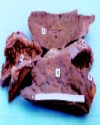
| Figures | Fig. 1:: Surface (a) and Cut Surface (d) view of portion of liver showing scarring, cystic bileduct (c) and the amphistomes (e)
|  | |
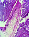
| | Fig. 2:: Photomicrograph showing amphistome in the bile duct along with mucosal proliferation, fibroplasia and cellular infiltration. H&E × 50
|  | |
|
| | |
|
|
|
|
║ Site map
║
Privacy Policy ║ Copyright ║ Terms & Conditions ║

|
|
|
742,751,723 visitor(s) since 30th May, 2005.
|
|
All rights reserved. Site designed and maintained by DIVA ENTERPRISES PVT. LTD..
|
|
Note: Please use Internet Explorer (6.0 or above). Some functionalities may not work in other browsers.
|